Knee Arthroscopic Surgery
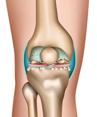
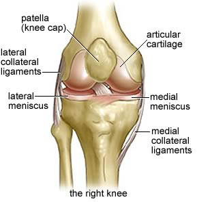
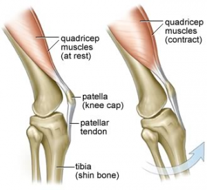
The knee joint is made up of three bones and a variety of ligaments. The knee is formed by the femur (the thigh bone), the tibia (the shin bone), and the patella (the kneecap). Several muscles and ligaments control the motion of the knee and protect it from damage at the same time. Two ligaments on either side of the knee, called the medial and lateral collateral ligaments, stabilize the knee from side-to-side.
Knee Arthroscopic Surgery
Arthroscopy is a minimally invasive ‘Key Hole’ surgical procedure on a joint in which an
endoscope inserted into the joint through a small incision for treatment of the injury. It is
technically possible to do an arthroscopic examination of almost every joint.
Some of the
advantages of an arthroscopy compared with traditional open surgery include:
- • Less post-operative pain
- • Faster recovery time
- • Quicker return to function
- • Lower risk of complications
Meniscus Surgery
Menisci are two crescent-shaped (half moon) discs between the thigh bone (femur) and the leg bone (tibia). They serve as shock absorbers and have several other important functions too.
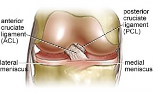
Ligament injury– ACL injury & Surgery
ACL stands for Anterior Cruciate Ligament of the knee.
There are two ligaments on the
sides of the knee: the Medial Collateral Ligament (MCL) and the Lateral Collateral Ligament
(LCL), and two crossed ligaments in the centre of the knee, the Anterior Cruciate Ligament (ACL)
and the Posterior Cruciate Ligament (PCL).
Cartilage Injury & surgery